


The human brain comprises about 87 billion neurons. On average, each of those cells makes hundreds of various connections to facilitate communication within the brain. Neural communication is believed to underlie all brain functions – from experiencing and interpreting the world around you, to remembering those experiences, to controlling your body's response.
But who exactly is talking to whom on this vast network of neural communication and what are the results of those individual conversations?
Understanding the small print of neural communication and the way it is formed by experience is considered one of the various focuses of neuroscience. However, that is complicated by the indisputable fact that there are such a lot of microscopic connections to check within the human brain, a lot of which are sometimes in flux, and that available tools cannot provide sufficient resolution.
As a result, many Scientists like me have turned to simpler organisms reminiscent of the fruit fly.
Scaplen et al. 2021/eLife, CC BY
Although fruit flies are annoying within the kitchen, they’re invaluable within the laboratory. Your brain is structured surprisingly just like that of humans. Importantly, scientists have developed tools that make studying fly brains much easier at resolutions not achieved in other organisms.
my colleague Gilead Barneaa neuroscientist at Brown University, and his team have spent over 20 years developing a tool that may visualize all of the microscopic connections between neurons within the brain.
Neurons communicate with one another by sending and receiving molecules called neurotransmitters between receptor proteins on their surface. Barneas Tool, tangotranslates the activation of specific receptor proteins into gene expression, which ultimately enables visualization.
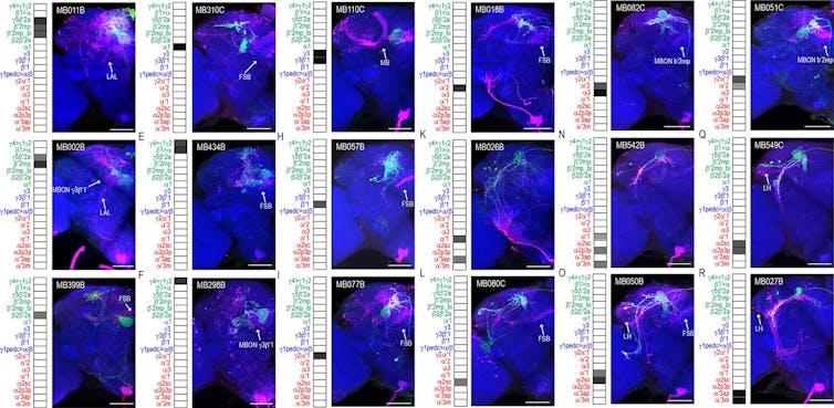
My team and I used -Tango to visualise all of the neural connections of a learning and memory center called “Tango”. Mushroom bodieswithin the brain of the fruit fly.
Kristin Scaplen, CC BY-SA
Here, a gaggle of about 4 neurons labeled in green receive messages from the mushroom body, the L-shaped structure with the blue label at the middle of the fly's brain. You can Step through the brain and see all the opposite neurons they’re likely communicating with highlighted in red. The cell bodies of neurons are situated at the perimeters of the brain, and the sites where they receive messages from the mushroom body appear as green tangles that enter a small oval compartment. Where these mesh-like green extensions mix with red, neurons are thought to transmit their processed message to other neurons downstream.
As you progress further into the brain, you possibly can see how the downstream neurons navigate to a single layer of a fan-shaped structure within the brain. The fan-shaped body It is believed to modulate many functions including excitement, Storage, locomotion And Transform sensory experiences into actions.
Our images not only revealed previously unknown connections throughout the brain, but additionally provide the chance to check the results of those individual neural conversations. The flies' brain connections were remarkably consistent, but additionally varied barely from one fly to a different. These subtle differences in connectivity are likely influenced by the fly's individual experiencessimilar to they’re with humans.
The great thing about tango lies in its flexibility. In addition to visualizing connections, scientists can use genes to control neural activity and higher understand how neural communication affects behavior. Because fly brains are structured similarly to human brains, researchers can use them to check how brain connections work and the way they may be disrupted in disease. Ultimately, this may improve our understanding of our own brains and the human condition.
image credit : theconversation.com