


Jena, Germany, 1924: They work almost in isolation and with meticulous boredom Psychiatrist Hans Berger observed rhythmic electrical activity on the scalp of human subjects. He is convinced that the activity originates within the brain and coins the term “electroencephalogram”.
It took 10 years for the scientific community to just accept Berger's work. Birth of the sphere of electroencephalographyEEG for brief.
Today the electroencephalogram – also abbreviated EEG – is is widely known As a Medical test to measure the electrical activity of the brain This is utilized in patients who’ve or are suspected of getting neurological disorders. EEG provides a glimpse into the living brain, providing a continuous electrical display of what is occurring inside our heads. The procedure may be short, often taking just half-hour. However, for patients being monitored to diagnose or treat a brain disease, treatment may proceed for for much longer – days and even weeks.
As a Neurologist specializing in epilepsyI exploit the EEG each day. Our team on the University of Florida interprets 1000’s of EEGs in neurological patients annually. Me too operate a research laboratory Our goal is to grasp the fundamental structure of the EEG in health and disease.
A story of unexpected turns
The history of EEG is colourful and stuffed with fables. Berger's interest in brain electricity was to not fight disease, although that was his essential job as a physician, but to search out a biological basis for his work Belief in telepathy. He wondered if EEG brain waves could transmit thoughts through space, allowing people to read one another's minds. Although his mission was unsuccessful, the sphere he founded still boomed.
In the mid-Nineteen Thirties, researchers observed the striking differences between waking and sleeping EEG. The EEGs of patients with brain diseases revealed a wide range of unprecedented observations.
And then one got here crucial moment for contemporary medicine. In December 1934, a gaggle of Boston physicians observed the occurrence of rhythmic EEG spike-wave seizures in patients with “Petit mal epilepsy. Petit Mal is an anachronistic term for a type of epilepsy wherein a patient's flow of thoughts, speech, or actions temporarily freezes during seizures. For the primary time, patients' symptoms and behavior during seizures have been correlated with a brain signal occurring in lockstep.
EEG quickly evolved from a scientific curiosity to a typical clinical tool. The first clinical EEG laboratory was established Massachusetts General Hospital in 1937. The practice evolved over the next many years into the specialized services that institutions like ours have been offering because the Nineteen Seventies.
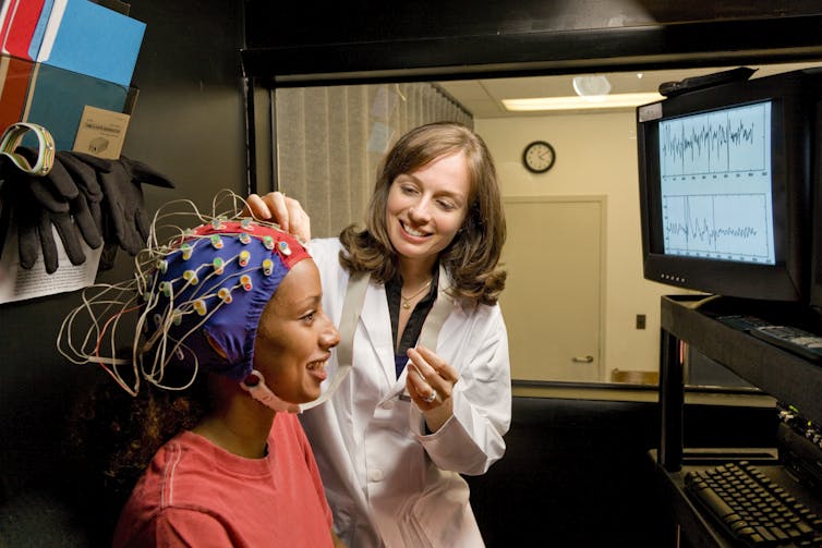
William Taufic/The Image Bank via Getty Images
The EEG explains
What exactly is the EEG?
Imagine taking two small metal disks connected by a conductive wire. Place one disc on the scalp and connect the opposite to a neutral reference, equivalent to the ear. Observe how a tiny alternating current flows within the wire, proportional to the electrical activity sensed by the conducting contact. This activity is the EEG, the electrical environment that bathes the brain tissue.
The EEG, in turn, is created by the excitability of nerve cells or neurons. When neurons fire, motion potentials—short, high-voltage spikes that travel outward from their cell bodies—trigger local electrical activity in other neurons, causing current to flow inside and out of doors of those neurons.
These local current flows could cause the goal neurons to fireside in turn and generate much more current flows. Thus, the system is self-sustaining. The overall average activity is a mix of many various frequencies, with the five essential frequencies called delta, theta, alpha, beta and gamma waves.
If the EEG were only a random drifting up and down – “the bloodless dance of action potentials,” he commented a skeptical neurologist of the early twentieth century – it could be much less interesting. The remarkable fact is that EEG tends to spontaneously organize into patterns in time and space.
The previously mentioned Petit Mal spike wave pattern is a classic example, but quite a few others at the moment are known. In clinical EEG practice, only characteristic EEG patterns are recognized and related to certain disease states.
Fluctuating neurons
Beyond the clinic, a troubling scientific query arises. Simply put: How do electrical patterns arise within the brain? How do the billions of neurons and their trillions of local current flows fluctuate in only the correct technique to create a globally ordered structure?
Our research group was taken with the basic query of pattern formation within the EEG. It seems that activity within the brain is inherently repetitive – that’s, oscillating. This is attributable to the best way neurons are connected to one another and the indisputable fact that they interact through excitation and inhibition To create push-pull effects.
By considering local oscillations as fundamental constructing blocks, we showed that EEG may very well be distributed throughout the brain built from such basic constructing blocks. Even more interesting is that different frequencies may very well be merged or synchronized into a typical rhythm. We have recognized that synchronization of this nature underlies some Seizure-like patterns have been observed in patients.
EEG, AI and the mind
Pattern formation in nature is deeply fascinating. How does a leopard get its spots? How do you get the audience to spontaneously burst into rhythmic applause at a concert? Many such questions have their origins in a classic work on biological patterns, published in 1952. Its creator was Alan Turinghigher referred to as the daddy of computer science and an early advocate of artificial intelligence, or AI.
The hardware underlying most of today's AI systems are neural networks. Neural networks were introduced in 1943 by Warren McCulloch, a physician and electroencephalographer. Like Berger, McCulloch's interest in EEG went beyond brain diseases. He wondered where the flexibility to think lies within the neurons and EEG of the brain. He had the thought of combining artificial neuron-like computing units into networks, analogous to the networking of real neurons within the brain.
Together with Walter Pitts, he proved that such neural networks could function as general-purpose computers. The groundbreaking McCulloch-Pitts ideas were refined over the next many years and are present in the “deep learning” neural networks of today’s AI.
Deep learning AI has infiltrated all areas of biomedicine, including neurology. For example, AI systems can successfully interpret brain scans. AI methods were also used Used for EEG evaluation.
Can AI systems be trained to derive thoughts from EEG? Can AI come closer to Berger's seek for telepathy? Incredible, current deep learning AI research has shown that some points of mental activity may be decoded from the EEG.
In 2024 the EEG shall be 100 years old. What windows will it open within the brain and mind in the long run? Undoubtedly, clinical applications will increase. The development of brain patterns will definitely be higher understood. Perhaps the EEG provides insight into the contents of the mind. And for neurologists like me watching the AI revolution, there may be a quiet pride that EEG really was at the start of all of it.
image credit : theconversation.com