


Cerebrospinal fluid or CSFis a transparent, colorless fluid that plays a vital role in maintaining the health and performance of your central nervous system. It cushions the brain and spinal cord, delivers nutrients, and removes waste products.
Despite their importance, problems related to cerebrospinal fluid often go unnoticed until something goes unsuitable.
I’m a Neurologist and headache specialist. In my work with patients with CSF pressure disorders, I actually have seen many alternative manifestations of those conditions. Here's what happens when your cerebrospinal fluid stops working:
What is cerebrospinal fluid?
CBF consists of Water, proteins, sugars, ions and neurotransmitters. It is principally produced by a network of cells that Choroid plexuswhich is positioned within the ventricles or cavities of the brain.
The choroid plexus produces approx. 500 milliliters of cerebrospinal fluid each daybut within the central nervous system only about 150 milliliters are present at anybody time, because the brain is always being absorbed and replenished. This fluid circulates through the Ventricles of the brainThe Central canal of the spinal cord and the Subarachnoid space across the brain and spinal cord.
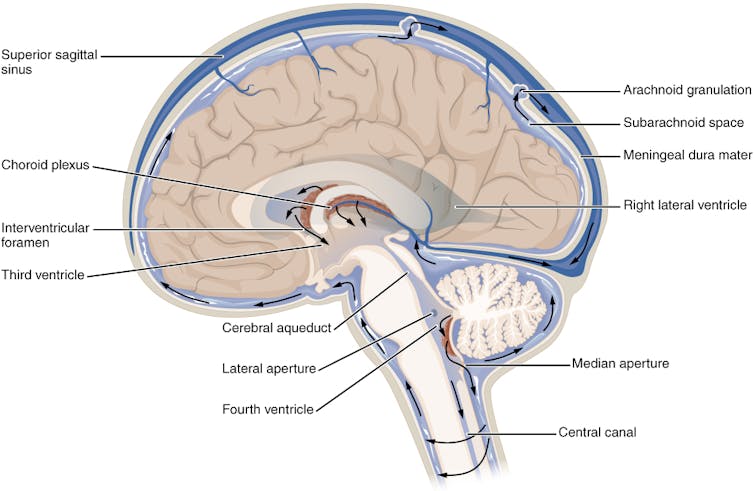
OpenStax, CC BY-SA
CSF has several critical functions. It protects the brain and spinal cord from injury by absorbing shock. Suspending the brain on this fluid reduces its effective weight and prevents it from being crushed by its own mass. In addition, CSF helps maintain a stable chemical environment within the central nervous system, facilitating the removal of metabolic waste and the distribution of nutrients and hormones.
When the production, circulation, or absorption of cerebrospinal fluid is impaired, it might probably cause significant health problems. Two necessary conditions are cerebrospinal fluid leaks and idiopathic intracranial hypertension.
Cerebrospinal fluid leak
A Cerebrospinal fluid leak occurs when fluid escapes through a tear or hole within the dura mater – the tough outermost layer. the meninges that surrounds the brain and spinal cord.
The dura will be damaged by head trauma or punctured during surgical procedures involving the sinuses, brain, or spine, corresponding to a lumbar puncture, epidural, spinal, or myelogram. Spontaneous CSF leaks can even occur with none identifiable cause.
Originally it was assumed that CSF leaks were relatively rare. Incidence of 5 per 100,000 peopleHowever, with increasing awareness and advances in imaging, healthcare providers are discovering increasingly more leaks. They are likely to occur more steadily in middle-aged adults and are more common in women than in men.
Risk aspects for the disease include connective tissue diseases corresponding to Ehlers-Danlos syndrome and postural orthostatic tachycardia syndrome.
Unfortunately, it is not uncommon for healthcare providers Misdiagnosis of a CSF leak than one other disease, corresponding to migraines, sinusitis or allergies. What can Diagnosis of a CSF leak difficult is the broad symptomatology. Most individuals with a CSF leak have a Positional headache This improves when lying down and gets worse when standing. The pain often occurs at the back of the top and might extend to the neck and between the shoulder blades. In addition to the headache, patients may experience ringing within the ears, visual disturbances, memory problems, difficulty concentrating, dizziness and nausea.
Imaging might help with diagnosisincluding an MRI of your brain or entire spine, or a myelogram of the space surrounding your spinal cord. Characteristics of a CSF leak which are visible in a scan are your Brain sags within the skull base and a fluid accumulation outside the dura. However, it’s estimated In 19% of patients with a CSF leak, the scans are normalsubsequently, it can’t be completely ruled out that there’s a leak if there are not any signs in the images.
Conservative treatment If you have got a CSF leak, you need to rest, lie flat, and drink more fluids to offer your spine time to heal the puncture. Increasing your caffeine intake to the equivalent of three to 4 cups of coffee a day can also help. increasing CSF production by stimulating the choroid plexus. Caffeine also relieves pain by interacting with Adenosine receptorswhich play a key role within the body's pain perception mechanisms.
If a conservative approach isn’t successful, Epidural blood patch could also be crucial. This procedure involves drawing blood out of your arm and injecting it into your spine. The injected blood might help form a covering over the outlet and promote the healing process. The headache may improve quickly, but when the patch doesn’t work or the outcomes are short-lived, additional tests could also be needed to raised locate the positioning of the leak. In rare cases Surgery could also be beneficialMost patients with a CSF leak reply to some form of those treatments.
Idiopathic intracranial hypertension
Idiopathic intracranial hypertension is a condition through which excess cerebrospinal fluid increases pressure within the skull and compresses the brain. The term “idiopathic” signifies that the reason behind the increased pressure is unknown.
Most patients with idiopathic intracranial hypertension have a History of Obesity or recent weight gain. Other risk aspects include taking certain medications corresponding to tetracycline, excessive vitamin A, tretinoin, steroids and growth hormones. Overweight middle-aged women are 20 times more likely Patients with idiopathic intracranial hypertension are diagnosed at a better rate than other patient groups. As obesity becomes more prevalent, the incidence of this disease can be increasing.
Patients with idiopathic intracranial hypertension typically experience Headaches and visual disturbancesTinnitus or eye pain. Papilledemaor swelling of the optic disc, is the standard finding in a Funduscopic examination the back of the attention. Doctors can also observe paralysis of the patient's eye muscles.
Brain imaging in patients with suspected idiopathic intracranial hypertension is crucial for Exclusion of other causes of increased cerebrospinal fluid pressure, corresponding to brain tumors or blood clots within the brain. Lumbar puncture or spinal puncture Measuring the pressure and composition of the cerebrospinal fluid can be crucial for diagnosis.
Since high intracranial pressure can damage the optic nerve and result in everlasting vision lossThe most important goal of treatment is to scale back pressure and preserve the optic nerve. Treatment options These include weight reduction, dietary changes and medications to scale back cerebrospinal fluid production. Surgical procedures can even reduce intracranial pressure.
Future directions and unknowns
Cerebrospinal fluid is important for brain health. Despite advances within the understanding of CSF-related diseases, some features remain unclear.
The exact mechanisms resulting in conditions corresponding to CSF leaks and idiopathic intracranial hypertension should not fully understood, but there are many theoriesFurther research is required to enhance diagnostic accuracy and supply more practical treatments for cerebrospinal fluid disorders.
image credit : theconversation.com